
NEWS
TM184C Builds Cell Bridges With a Receptor-Like Fold
Miami scientists found TM184C, a GPCR-like vesicle protein that builds cell bridges, ships organelles, and holds back autophagy.

A Miami team found TM184C, a GPCR-like protein that builds cell bridges and limits autophagy, by matching shapes across 214,528,851 AlphaFold models. Kyutae D. Lee, Samuel Taylor and Jennifer Arcuri share first authorship on the open-access study published on 9 September, with Daniel G. Isom as senior author at the University of Miami Miller School of Medicine and Sylvester Comprehensive Cancer Center.
The protein looks like the receptors that dominate drug lists. It lives on moving vesicles, strings cells together with thin tubes, and holds back the cell’s recycling programme. Sequence search had left it in the dark.
A Shape Search of 214 Million Proteins
Most protein names still come from matching letters in a sequence. Isom’s group asked what is missed when the letters no longer match but the fold still does. They define those cases as superdark: structural cousins of known proteins that lack a clean sequence hit, a bin darker than the already poorly mapped Tdark slice of the human proteome, which covers 5,516 of 20,412 proteins (27%).
On 20 July 2023 they pulled AlphaFold2 version 4 onto the Frost Institute clusters at Miami. The compressed archive occupied 24 TiB. They then ran TM-align against the rhodopsin structure used as the search query, the classic seven-helix GPCR from cattle eye.
THE SHAPE SEARCH
| Filter | Count |
|---|---|
| AlphaFold2 models searched | 214,528,851 |
| TM-align hits (at least 200 residues, score 0.5) | 1,543,898 |
| After obsolete UniProt IDs removed | 1,461,479 |
| Rank 1 / 2 / 3 fold quality | 47% / 36% / 17% |
| Known GPCRs | 506,129 (35%) in 15,368 organisms |
| Non-GPCR seven-helix proteins | 955,350 (65%) in 371,568 organisms |
| Human superdark families kept | TM184 and PRRT (6 proteins) |
A sequence network of 39,808 trimmed seven-helix cores then linked archaeal histidine kinases to eukaryotic GPCRs in at most six BLAST steps, using a 30% identity floor checked against 2,029 human models. The human list started at eight names. TMEM116 and TMEM187 were dropped because they had already been floated as GPCRs. Six remained: TM184A, TM184B, TM184C, SLC51A, PRRT3 and PRRT4.
Among more than 800 human GPCRs, more than 100 still sit in Tdark. TM184C was the broadest of the new set on Human Protein Atlas charts, so the wet lab followed it. UniProt’s 438-amino-acid TMEM184C protein record still carries an older line, “possible tumour suppressor,” from a 2007 anaplastic thyroid growth assay. Pharos still tags it Tdark.
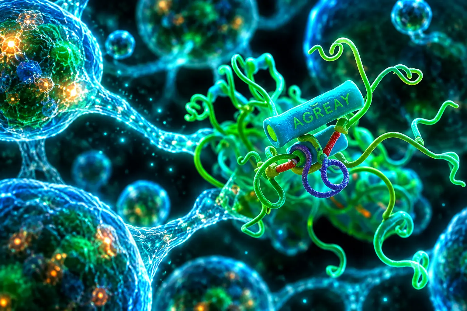
TM184C Recruits Arrestin Without G Proteins
The fold is seven helices. The biochemistry is only half familiar. TM184C lacks the classic class A switches (LxxD, CWxP, PIF, NPxxY, E/DRY). It does carry an NRY triplet at N135-R136-Y137, lined up with rhodopsin’s E134-R135-Y136, and a glutamine in helix 6 that would break the usual ionic lock.
What it does have, in abundance, are C-terminal arrestin codes, clusters of serines and threonines that GPCR kinases phosphorylate so β-arrestin can bind. TM184C has four of those codes. TM184A has one, TM184B two, PRRT3 and PRRT4 six. Mass spectrometry mapped phosphates on S422, S432 and S435. An arrestin-code mutant that changed those three plus eight neighbouring sites (S374, S377, S379, S380, S382, S424, S427, S438) to alanine pulled down less ARRB1. Cutting the last 82 residues (TM184C-Δ82) did the same.
GRK2, GRK3, GRK5 and GRK6 all increased arrestin recruitment, GRK2 most of all, and GRK2 bound full-length TM184C as strongly as it bound the β2-adrenergic receptor ADRB2. Lambda phosphatase collapsed the upper band of a TM184C doublet, confirming the shift was phosphate. In 4A, mini-G and TRUPATH assays, the same protein did not recruit active-state G proteins the way ADRB2 or MRGPRD did. A small Gs dip in TRUPATH did not stay above background.
For decades, we have largely explored protein biology using sequence as our guide. We wanted to know what biology we might be missing if we searched by three-dimensional structure instead. What we found suggests there is another layer of biology that has been hiding in plain sight.
Daniel G. Isom, molecular pharmacologist, University of Miami
The authors liken the pattern to atypical chemokine receptors, which also lean on arrestin rather than G proteins. Bystander BRET put TM184C in late endosomes and lysosomes, with almost no plasma-membrane signal, while TM184A and TM184B reached the cell surface.
Vesicles That Build Bridges Between Cells
Live imaging in HEK293A cells put TM184C-eGFP on vesicles that run both ways along microtubules, splitting and fusing as they go, and that co-localise with the kinesin KIF5B. Some vesicles are acidic on LysoTracker; some are not. LC3B-mCherry rides with them, so autophagosomes are in the mix. Short projections packed with these vesicles measured about 5 micrometres and 23 micrometres. Longer ones grew from about 50 micrometres to 58 micrometres in 3 seconds.
Those arms fuse. The resulting bridges look like tunnelling nanotubes and tumour microtubes, and TM184C vesicles pile up at the tips where two arms meet. In established bridges the vesicles line the tube and wrap acidic organelles, a layout the authors read as a route for lysosomes and other cargo to change cells.
Jennifer Arcuri, a co-first author and Sylvester scientist, said the moving vesicles forced a rewrite of the protein’s job. “When we saw TM184C-positive vesicles moving through connections between cells, we realized these structures could be routes for substantial material exchange,” she said. “That completely changed how we thought about TM184C and made us consider how cells might use these connections to cooperate and compete for resources.” Shraddha Chandthakuri, a cancer-biology graduate student in the Isom lab, put the tension in one line: “I think cells coordinate until they have to compete.”
KNOCKDOWN VERSUS OVEREXPRESSION
- CRISPR loss: HEK293A and T cells rounded up, dropped projections and bridges, and filled with enlarged, mis-placed acidic vesicles; they blebbed and died under the imaging laser.
- Stable excess: TM184C-eGFP or TM184C-mCherry cells grew arms spanning tens to hundreds of micrometres and scored far higher on a bridge-projection-protrusion (bpp) index (n = 28-60 regions per condition).
- Rescue test: Recoded full-length TM184C kept the high-connectivity look after endogenous TM184C was cut; the ACM and Δ82 tails did not.
- Cargo flow: TM184C-to-TM184C co-cultures swapped vesicles both ways; TM184A, TM184B and ACM cells mostly received cargo; Δ82 “transfer” was mislocalised protein riding degradative vesicles.
The same TM184C-positive tubes and vesicle swaps appeared in HFF-1 fibroblasts, astrocytes, SK-MEL-28 melanoma cells, and mixed cultures of astrocytes with patient-derived glioblastoma cells. TM184C vesicles tracked mitochondria in kiss-and-go contacts inside HEK293A cells, astrocytes and GBM cells, including along the bridges.
Isom’s laboratory posted the paper on 9 September and framed it as a look into a protein universe whose named members are still a minority.
Out online today in @Nature. The known protein universe holds hundreds of millions of members, yet most remain unexplored, a vast biological darkness that may hold important clues to human health and disease. We went looking in it. https://t.co/EkE584rOQL pic.twitter.com/PUyHEehgUe
— IsomLab (@danIsomLab) September 9, 2026
Every author is at the University of Miami. High-performance computing came from Warner Baringer and Elvis Maradzike at the Frost Institute. Funding listed on the thread includes an NIH R35 award from NIGMS, Sylvester, and Isom’s Pap Corps endowed chair.
Why Glioblastoma Networks Look Familiar
Glioblastoma cells are famous for tumour microtubes, open cytoplasmic channels that turn a tumour into a network and help it share damage and resist treatment. An April 2026 AACR abstract from Jennifer Arcuri, Bruno Colon, Chandthakuri and Isom stated that TM184C expression correlates with poor prognosis in GBM, and that HEK293A cells overexpressing the protein grow connections that phenocopy TM184C-positive structures in patient-derived GBM cells. Median survival for GBM remains about 20 months in that abstract’s framing.
The Nature paper itself does not run a survival analysis. It does show TM184C vesicles and mitochondrial contacts in patient-derived GBM cells, and it shows that the C-terminal arrestin code is required for productive vesicle transfer. That is enough to put the protein on the same map as tumour microtubes, not enough to say it causes the bad outcomes.
WHAT WE KNOW
- In culture: TM184C is required for TNT-like bridges and for bidirectional vesicle exchange in HEK293A cells.
- In GBM cells: Patient-derived GBM cells form TM184C-positive connections and keep TM184C vesicles close to mitochondria.
- In yeast: Human TM184C restores vacuole shape after HFL1 deletion; TM184A and TM184B do not.
WHAT IS UNCONFIRMED
- Ligand: No endogenous or synthetic binder is assigned.
- G protein: Weak or tissue-specific coupling is not ruled out, only not seen in the assays used.
- GBM causality: The poor-prognosis correlation is stated in the AACR abstract; a clinical series is not in the Nature figures.
UniProt’s older growth-suppressor note and the new network-builder phenotype can both be true in different assays. A protein that builds bridges can still slow a dish of thyroid cells when it is forced on, and still help a brain tumour share mitochondria when it is left in place. Those are different experiments, not a single verdict.
Yeast Vacuoles Still Run on the Human Protein
Sequence traces the TM184 family to archaea, which is why the authors treat the autophagy role as older than multicellular life. The yeast homologue is Hfl1, a vacuole-membrane protein named Has Fused Lysosomes. A 2018 crystal study showed that Hfl1 recruits yeast Atg8 to vacuoles through a helical AIM, not the usual beta-strand Atg8 motif, and that this binding supports lipidation-independent vacuole shape.
Deleting HFL1 in Saccharomyces cerevisiae gave the multivesicular vacuole look that marks broken autophagic-body handling. CRISPR knock-in of human TM184C fully restored that morphology. TM184A and TM184B did not. An antibody raised against human TM184C recognised the knock-in protein on yeast blots, with porin as the load control.
THE YEAST RESCUE
| Protein | Home membrane | Autophagy readout | Bridge / transfer |
|---|---|---|---|
| TM184C | Late endosomes, lysosomes, autophagosomes | Limits LC3B-II; knockdown raises autophagosome formation | Required for TNT-like bridges and two-way vesicle flow |
| Hfl1 | Yeast vacuole membrane | Autophagic-body homeostasis; binds Atg8 | Not a multicellular bridge protein |
| TM184A / TM184B | More at the plasma membrane | Did not rescue hfl1-null vacuoles | Mostly receive TM184C cargo, do not drive it |
In HEK293T cells, CRISPR knockdown of TM184C stacked up lipidated LC3B-II. Concanamycin A at 1 nM for 18-24 hours pushed LC3B-II higher still, which the authors read as extra autophagosome birth, not a blocked lysosome. Bafilomycin A1 did the same and left cells full of enlarged TM184C- and LC3B-positive vesicles. TM184C therefore acts as a brake on autophagosome build-up, a job the human protein can still do on a yeast vacuole.
What a 7-Helix Housekeeper Means for Drugs
GPCRs are the industry’s favourite membrane proteins because chemists already know how to work a seven-helix bundle. A 2025 survey of the class counted 516 approved drugs targeting GPCRs, 36% of all approved drugs, acting at 121 targets, with 337 further agents on 133 GPCRs including 30 novel ones. Diabetes and obesity medicines in that class booked nearly $30 billion in 2023 sales.
GPCR MEDICINES IN 2025
- Approved set: 516 GPCR drugs, 36% of all approved drugs, 121 targets.
- Pipeline: 337 agents on 133 GPCRs, 30 of them novel targets.
- Still dark: More than 100 human GPCRs remain Tdark; TM184C was not even on that GPCR list until the shape search.
- Metabolic cash: Nearly $30 billion in 2023 sales for diabetes and obesity GPCR drugs.
TM184C is not a new surface receptor waiting for a hormone. It is a vesicle protein that uses the same C-terminal phosphate-and-arrestin logic to decide where cargo goes, including across a bridge into a neighbour. That is a second job for a fold the industry already screens, and it cuts two ways. A compound aimed at “a GPCR” could hit a trafficking protein by mistake. A compound aimed at tumour microtubes or at autophagy now has a seven-helix handle, if anyone can find a ligand or an arrestin-biased probe.
Isom was blunt about the method. “AI cannot be blindly trusted, but can lead to really big things in the hands of experts and prepared minds,” he said. The paper’s own next steps are ordinary and hard: a real structure of TM184C, a ligand if one exists, and a clean test of whether cutting the bridges in a tumour is help or harm. Until those land, the result that is already on the table is simpler. A housekeeper with a receptor’s body was sitting in the AlphaFold pile, and yeast still runs on the human version.
Frequently Asked Questions
What Gene Encodes the TM184C Protein?
The human gene is TMEM184C, also catalogued as TMEM34 and PRO1355, on chromosome 4. The canonical chain is 438 amino acids and 50,142 daltons. UniProt lists a second splice form that lacks residues 262-438. Akaishi and colleagues reported in 2007 that the transcript was down in 11 anaplastic thyroid cancer lines and that forcing it slowed KTA2 cells, which is why the database still mentions a possible tumour-suppressor role.
What Does Superdark Mean in the Miami Study?
Superdark, as the authors use it, means a protein that matches a known fold in three dimensions while lacking a definitive sequence match to that family. The Illuminating the Druggable Genome scheme already splits human proteins into Tclin, Tchem, Tbio and Tdark; superdark sits beyond Tdark. The search returned two such human families, TM184 and PRRT.
Does TM184C Couple to G Proteins?
Not in the assays reported. 4A, mini-G and TRUPATH readouts that lit up ADRB2 or MRGPRD stayed at background for TM184C, and a small Gs TRUPATH change was not reproducibly above noise. The authors leave room for weak or context-dependent coupling they did not capture. Arrestin binding and GRK phosphorylation, by contrast, were robust and mapped to the C-terminal tail.
How Did Knockdown Change Autophagy?
Losing TM184C raised the lipidated LC3B-II band on HEK293T blots. A further rise after 1 nM concanamycin A for 18-24 hours told the authors that autophagosomes were still being cleared, so the primary defect was extra formation rather than a lysosomal jam. Bafilomycin A1 produced the same pile-up of enlarged TM184C- and LC3B-positive vesicles on the microscope.
What Is Hfl1?
Hfl1 is the yeast protein in the TMEM184 family (InterPro IPR005178). The name stands for Has Fused Lysosomes. Liu and colleagues showed in 2018 that it binds Atg8 through a helical AIM and that this binding supports vacuole shape without Atg8 lipidation. The Miami knock-in shows that human TM184C, and not TM184A or TM184B, can stand in for that yeast job.
Disclaimer: This article is news reporting on laboratory research and is for information only. It is not medical advice and does not recommend any test, drug, or treatment for glioblastoma, autophagy-related disease, or any other condition. Readers who need clinical guidance should speak with an oncologist or another qualified physician before making health decisions. Protein functions, expression links, and drug-class figures come from the cited papers and databases and may change as other laboratories repeat or extend the work.

The BMW iX4 Undercuts Mercedes and Trails Its Twin

Scotland’s Ten Big Supermarkets Already Hold the Weekly Shop

Scotland EMS Faces a State Probe After a Wagram Death

Meritz Signs Ripple to Race Korea’s 2027 Token Rules

Pocognoli’s First Camp Leaves Scotland Third and Exposed

Volkswagen Sells the Osnabruck Plant to Clear a Board Fight

Saksham Gaur Finds He Is His Own Company’s Client

Tata Motors’ Iveco Tender Opens as Holders Still Decide

Onimusha Way of the Sword Preload Hits a Windows 11 Wall

Apple’s Foldable iPhone Hinges on Samsung Display Supply
-
 AUTO1 month ago
Volkswagen Sells the Osnabruck Plant to Clear a Board Fight
-
 BUSINESS2 months ago
Saksham Gaur Finds He Is His Own Company’s Client
-
 AUTO2 months ago
Tata Motors’ Iveco Tender Opens as Holders Still Decide
-
 GAMING2 months ago
Onimusha Way of the Sword Preload Hits a Windows 11 Wall
-
 NEWS2 months ago
Apple’s Foldable iPhone Hinges on Samsung Display Supply
-

 AUTO2 months ago
Simple Wave Chases Range as Ather Konarc Cuts Price
-

 NEWS1 month ago
The Frame Pro’s $1,200 Labor Day Cut Needs a Soundbar
-

 NEWS1 month ago
iQOO 16 Locks 8,400 mAh While 100W Charging Holds