Edinburgh has officially joined the elite club of cities equipped with a total-body PET scanner, placing Scotland at the heart of the UK’s most advanced medical imaging network to date. It’s not just a technological milestone — it could rewrite how fast we detect disease, test new drugs, and treat patients.
Located at the Royal Infirmary of Edinburgh, the scanner is now operational under a cross-institutional effort co-managed by the Universities of Edinburgh and Glasgow. The move marks a significant leap forward for NHS Scotland and brings game-changing imaging capabilities beyond the M25 for the first time.
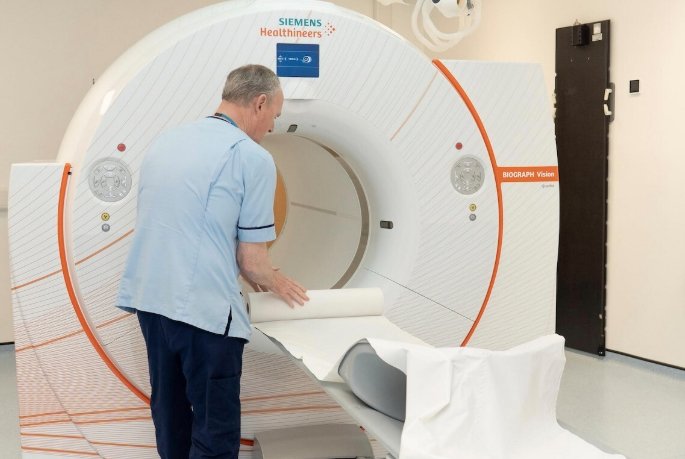
A Scanner That Sees Everything, Everywhere, All at Once
The Biograph Vision Quadra PET/CT system, built by Siemens Healthineers, is not your typical hospital scanner. It doesn’t just look inside you. It captures nearly your whole body — simultaneously, in seconds, in vivid detail.
Unlike standard PET machines that scan a slice at a time, the total-body PET scanner is about 40 times more sensitive, delivers 10 times faster imaging, and can scan up to 50% more patients each day. That’s not an upgrade — that’s a transformation.
And Edinburgh’s not alone anymore. London already has two — one under the NPIP scheme at St Thomas’ Hospital (live since late 2024), and another privately funded at Royal Free Hospital. Now Scotland is in the mix.
One-liner impact: Scotland now sees what the body’s doing, in real-time, with a clarity most of the world still dreams about.
National Imaging Grid, Global Implications
This isn’t just a scanner dropped into a hospital. It’s part of a much bigger vision — the National PET Imaging Platform (NPIP) — funded by £32 million through the UKRI Infrastructure Fund and backed by heavyweight organisations like the Medical Research Council and Innovate UK.
The platform aims to:
-
Standardise PET imaging across the UK
-
Open data-sharing channels for clinical trials
-
Accelerate drug development
-
Enable AI training at scale
“Researchers gain not just better data — they gain connected data,” said Dr Juliana Maynard, NPIP’s Director of Operations. “From here on out, the research and clinical ecosystems are no longer siloed.”
And that means a company testing a cancer drug in London can now extend trials seamlessly into Edinburgh, pulling clinical-grade imaging data across cities and studies like never before.
Why PET Is Such a Big Deal for Modern Medicine
If you’ve never had a PET scan, here’s why scientists are excited. It doesn’t just show a picture of your insides like an X-ray or CT.
It shows how your body functions — in real-time.
PET, or Positron Emission Tomography, reveals how tissues behave metabolically. In oncology, for example, it lights up aggressive cancers long before a lump is visible. In neurology, it shows chemical imbalances linked to Alzheimer’s or Parkinson’s. And in cardiology, it maps blood flow and tissue death post-heart attack.
Here’s how it works in a nutshell:
| Feature | Function |
|---|---|
| Tracer Used | Fluorodeoxyglucose (FDG), tagged with fluorine-18 |
| Detection Mechanism | Gamma rays from positron-electron annihilation |
| Imaging Type | Functional — shows glucose use, receptor binding, etc. |
| Time Sensitivity | Tracer decays fast (half-life: 109 minutes) |
| Applications | Oncology, neurology, cardiac disease, inflammation |
Basically, it’s like having a front-row seat to your cells in action.
A Giant Leap for Drug Development
For pharmaceutical companies and clinical researchers, the implications are massive. Access to real-time biological data during trials lets scientists see how an experimental drug distributes, binds, and acts — all in a single scan session.
This data helps answer the all-important “go or no-go” question faster than ever.
Professor Chris Molloy, CEO of Medicines Discovery Catapult, called it “a national capability for testing new treatments and enabling faster patient benefit.”
Key benefits for R&D include:
-
Real-time pharmacokinetics
-
Early proof of drug mechanism
-
Faster clinical go/no-go decisions
-
Reliable biomarker validation
And with a national PET imaging repository coming soon, all collected data — anonymized and standardized — will be pooled into a shared database. It’s Google Maps for molecular imaging.
From Physics Breakthrough to Everyday NHS Tool
It’s hard to overstate how far PET has come. The first machine for human use was installed in the early 1970s in Pennsylvania. The tech owes much to Michel Ter-Pogossian’s work in St. Louis and was developed further by Michael Phelps, often called the father of clinical PET.
Today, it’s not just an academic plaything. It’s part of the NHS toolkit.
Edinburgh’s machine, while capable of cutting-edge research, will also scan real patients — helping diagnose cancers, inflammatory diseases, and neurological disorders faster and more precisely.
“The NHS gains both diagnostic speed and accuracy,” one Edinburgh-based consultant radiologist told us. “For patients, that can mean earlier treatment. For doctors, more confidence in clinical decisions.”
The UK’s Global Play in Precision Imaging
Zoom out, and this is part of a bigger strategic bet. The UK wants to be seen not just as a place for great hospitals or great research labs — but the world’s best environment for testing and validating new medicines.
By planting total-body PET scanners in major cities and linking them into a nationwide platform, it becomes easier to:
-
Attract pharma trials from overseas
-
Win AI biotech investment
-
Lead in radiopharmaceutical development
“The network we’re building now wasn’t imaginable even 10 years ago,” said Maynard. “It’s both a national asset and a global signal.”
And the signal is loud and clear: Britain is serious about being a world leader in precision diagnostics and next-generation drug discovery.
 Windows 11 KB5095093 Lands With Point-in-Time Restore and 35-Day Pause
  Meta Cuts Smart Glasses to $299 to Outflank Snap and Apple
  Messi Leads the 2026 World Cup Golden Boot Race After Two Matches
  Australia March On as Athapaththu Ton Lights Up T20 World Cup
  Galaxy Z Fold 8 Leak Reveals Colors, Storage, and a New Ultra Variant
  Ronaldo’s Six-World-Cup Record Leads a Matchday Full of Milestones